


Many scientific imaging techniques or microscopes are available for life science, materials science, and engineering research. Each has its strengths and limitations. When you start new research or face a new problem to solve, you might grab the closest instrument available, but that might not be the best choice.
We can categorize imaging techniques by their abilities and related characteristics, such as:
- Resolution
- Applicable sample types and sizes
- Imaging dimensions (surface or volume, 2D or 3D)
- Destructive or non-destructive
- Obtained information (color, density, composition)
- Required sample preparation
- Measurement speed
Or, we can categorize them by the technologies used and related characteristics, such as:
- Source of illumination (light, electron beam, X-rays, neutrons)
- Type of magnification (optical, mechanical, digital, etc.)
- Instrument size
- Instrument cost
X-ray micro CT (computed tomography) is the technique I am most familiar with. It is used to obtain 3D images of samples with micron resolution. Over the years, I have consulted with and helped scientists and engineers choose the right technique or correlate multiple techniques.
In this article, I will review the concept of resolution, one of the most critical characteristics of a microscope. Then, I will compare three imaging techniques with 3D imaging capability and micron-scale resolution: micro-CT, scanning electron microscopy (SEM), and confocal laser scanning microscopy (CLSM).
For a more comprehensive list, I recommend Microscopy: A Very Short Introduction by Terence Allen, the Honorary Professor of Structural Cell Biology at the University of Manchester. It is a concise introduction to microscopy but covers an extensive list of techniques.
Resolution
What is resolution? “Resolution is the ability of the measurement system to detect and faithfully indicate small changes in the characteristic of the measurement result,” according to the NIST/SEMATECH e-Handbook of Statistical Methods. In imaging, “the small change in the characteristic of the measurement result” is the contrast change.
Here is a simple alternative way to describe it. Spatial resolution is the size of the smallest feature you can observe in the image because of the contrast difference between the feature and its surroundings.
Resolution is important because, if you use an instrument with insufficient resolution, you can’t see what you need to see.
The figure below gives you an idea of the resolution and instrument you need to see different things.
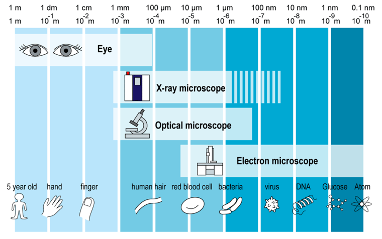
Depending on your eyesight, you can recognize things on a millimeter scale without magnification. You can use a magnifying glass to magnify an image by four or five times. Optical microscopes can increase the magnification factor to two thousand by combining lenses. Electron microscopes can increase it to about one million.
You need an X-ray or optical microscope to see things with a submillimeter or micron resolution, such as the surface texture of human hair. To see even smaller things, such as viruses and atoms, you need an electron microscope.
We need different techniques for each resolution range for two primary reasons:
- The theoretical resolution is limited by Abbe’s equation, which states that resolving power equals half the wavelength of the light used divided by the numerical aperture of the objective lens. (Diffraction limit)
- Available lenses and mechanical misalignment of instruments reduce the practically achievable resolution.
Reason 1 means that the wavelength of the readily available light fundamentally limits the resolution you can achieve. This is one reason - in addition to the availability of lenses - why electron microscopes can achieve high resolution. For example, you can generate an electron beam with a wavelength of 7 picometers at 30 keV.
Reason 2 means that you need a way to magnify images. This is where X-ray CT is limited despite its short wavelength. It is challenging to use lenses for X-rays. The refraction index of X-rays is close to 1, so they don’t refract much. We need special optics - such as super-mirrors and Fresnel zone plates - to “bend” X-rays.
This article goes into more detail about X-ray CT resolution:
Read: How to Improve the Resolution of X-ray CT Images
Now let’s compare CT, CLSM, and SEM.
X-ray micro-CT
How does it work?
Different materials have different absorption rates for a given X-ray energy, so the radiograph or intensity distribution of X-rays that pass through an object contains information about the density distribution in the object.
However, one radiograph is a 2D projection of three-dimensional density distribution. To obtain the 3D density distribution, which is the CT volume data, we need to rotate the sample to collect multiple projections and reconstruct a 3D volume.
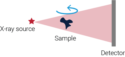
The most commonly used magnification method, called cone beam geometry, uses the divergence of the X-ray beam originating from a micron-size source, as shown in the figure above.
Read: What is micro-CT?
Advantages
X-rays can penetrate a wide variety of samples, so X-ray CT can provide their complete 3D images, including the internal structure, non-destructively. Any samples from a few to several hundred millimeters in size can be scanned. This is the biggest advantage of this technique.
Disadvantages
As mentioned above, the resolution is limited to a few microns to the submicron level for most laboratory CT scanners. It is limited to about 10 nm even with specialized nano-CT systems or synchrotron radiation.
- Example of 100 nm resolution using mirrors: Matsuyama et al., 2016, Sci. Rep., 6:24801
- Example of 10 nm resolution using Fresnel zone plates: De Andrade et al., 2021, Adv. Mater., 33(21), 2008653
Another drawback is that you can only see contrast if there is enough density contrast in the sample. This disadvantage can be overcome to some extent by staining samples or using low-energy and monochromatic X-rays.
Samples can also be too dense or X-ray-absorbing to image. In that case, neutron imaging can be an alternative technique.
Recommended applications
- Non-destructive testing of objects’ internal structures, including cracks, voids, and inclusions
- Studying materials’ microstructures, including particle, void, and fiber shape, size, and orientation analyses
SEM (Scanning Electron Microscopy)
How does it work?
SEM uses an electron beam as the source of illumination instead of X-rays or light. As shown in the figure below, the electron beam produced by an electron gun travels vertically through lenses and scan coils before reaching the sample. The entire path is in vacuum because air or gas can immediately absorb the electron beam.
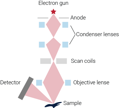
The scan coils control the electron beam position on the sample. Various interactions between the electron beam and the sample provide information about the sample. These interactions include secondary electrons (SE), reflected or back-scattered electrons (BSE), characteristic X-rays (energy-dispersive X-ray spectroscopy, EDS) and light (cathodoluminescence) (CL), absorbed current (specimen current), and transmitted electrons (TEM). Among these interactions, SEM uses secondary electrons.
SEM images only the near-surface area of the sample. You can use a focused ion beam (FIB) to slice the sample layer by layer while running an SEM scan for each layer. This technique is called FIB-SEM and can produce 3D data of your sample.
Watch An Introduction to Scanning Electron Microscopy and Focused Ion Beam by Dr. Matthew Bresin
Advantages
The biggest advantage of SEM is the resolution. SEM can typically achieve nanometer - order resolution because of the easily achievable short wavelength and availability of lenses.
Using specialized EDS and WDS (wavelength dispersive spectroscopy) detectors and software, elemental concentrations, mapping, mineral identification, and modal analysis can be collected using an SEM. These techniques are widely used in geology and metallurgy.
Disadvantages
Because traditional SEMs require the sample to stay in a vacuum and interact with an electron beam, some samples require special preparation. For example, live or wet cells need to be dried, or a non-conductive sample needs to be coated with a conductive material such as gold.
However, modern SEMs allow water vapor introduced into the chamber to allow non-conductive and wet samples to be imaged, typically at low kV settings.
You might think SEM is expensive, but the price starts from around $75K. This means some are less expensive or similar to micro-CT scanners.
Read: CT vs. SEM: Which Is Better?
Recommended applications
- Nanometer to micron resolution imaging of inorganic materials
- Semiconductor and microchip assembly inspection
- Cell morphology, microbiology, including investigation of the interactions of bacteria and viruses’ with surfaces
CLSM (Confocal Laser Scanning Microscopy)
How does it work?
Confocal microscopy was invented to overcome the drawbacks of fluorescence microscopy: photobleaching of the sample and blurring of fluorescence microscopy images. These problems were solved by changing the light source from a lamp to a laser and placing a pinhole in front of the detector.
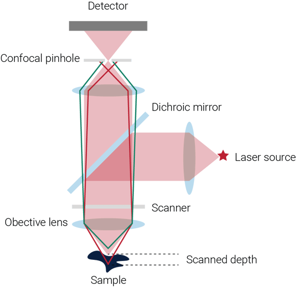
As shown in the figure below, the lasers reflected by the dichroic mirror focus on a small region of the sample. Then the sample emits fluorescent light, which is captured by the detector. The scanner, composed of two mirrors, changes the direction of the laser to hit different positions on the sample while each spot’s fluorescence response, which generates a sharp image when all images are combined.
The fluorescent light originating from different focal planes focuses on different positions around the pinhole. By adjusting the pinhole settings, you can choose a specific focal plane to scan and image. Repeating this process for multiple focal planes constructs a 3D image of the sample.
Advantages
The most significant advantage of CLSM is that it can image live cells and tissues. CLSM can also achieve about 200 nm resolution, which is better than micro-CT scanners, although not as good as SEM.
CLSM is relatively inexpensive. The price ranges from $10K to $100K.
Disadvantages
The drawback of CLSM is the limited depth you can image. The penetration depth of CLSM is limited to about 100 microns.
Recommended applications
- Imaging of cellular and soft tissue structures
- Studying cellular and molecular events, including in vivo experiments
Takeaway
The choice of imaging technique will depend on the specific application, sample type, and research aims. Some techniques might not be applicable to your sample, and you have only one choice. Or, when more than one technique can be used, comparing typical examples of three techniques helps you choose the best technique.
Sometimes, there is no one right technique, and combining multiple complementary techniques might be the best solution.
If you are wondering if CT is the right technique for your research, our team of CT experts can help you. You can talk to one of our CT experts by clicking the “Talk to an expert” button at the top right of the page or send us a message at imaging@rigaku.com.