X-ray micro-CT (computed tomography) is a powerful tool that allows researchers to look inside batteries in a non-destructive way. Swelling and delamination after repeated use can be visualized and quantified without opening the packages.
Analysis procedure
In this example, lithium-ion battery packs were scanned using a micro-CT scanner, CT Lab HX.
The CT images of the normal and damaged batteries were compared and inspected for damages.
The interior image of the damaged battery was segmented to identify battery components and delamination using deep learning segmentation. The thickness distribution of the delamination was calculated.
1. CT scan
Two lithium-ion battery packs, normal and damaged, were scanned to produce the 3D grayscale CT images.
2. Battery inspection
2D cross-sections for the two batteries are shown. The one labeled "damaged" shows signs of swelling and delamination within the battery layers. An intensity plot taken across each battery shows significant nonuniform spacing between layers in the ‘damaged’ battery.
2D cross-sections along the positive tab are shown in the figure. Both the normal functioning and damaged batteries show deflected anodes.
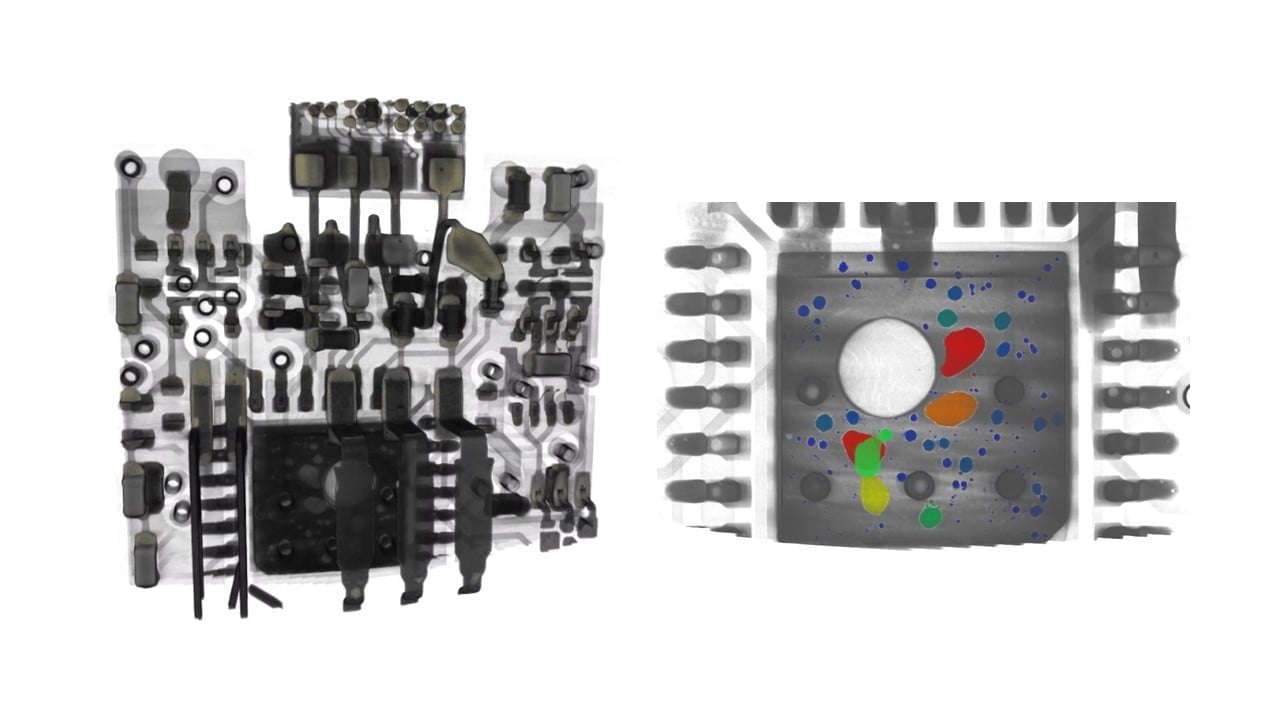
3. Segmentation and quantitative analysis of the delamination
Deep learning segmentation was performed for the CT image. The cathode is shown in blue and the anode is red. Areas of delamination can be seen in the nonuniform layers as darkened areas.
The size distribution of the delamination was calculated. The green and yellow areas indicate over 200 microns wide delamination.