X-ray Micro Computed Tomography Seminar and Workshop
In this seminar, we learned about various applications of X-ray micro-computed tomography (CT) in geology and archeology from the top researchers making the most of X-ray CT in their fields.

X-ray CT Menu

- Program
- Presenters
- Location
- Recording
Seminar and workshop program
8:30AM - 9:00AM
Registration (Room ZHS 265)
30 minutes
9:00AM - 9:15AM
Opening Remarks (Room ZHS 265)
15 minutes
9:15AM - 10:00AM
Integrating microCT to improve research and pedagogy in Earth Sciences (Room ZHS 265)
45 minutes
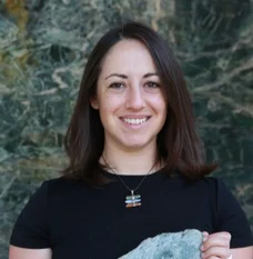
Dr. Emily Cooperdock
Assistant Professor, Department of Earth, Environmental & Planetary Sciences
Brown University
10:00AM - 10:45AM
Assessing trabecular bone architecture at the divergence of bird-line (Avemetatarsalia) and croc-line (Pseudosuchia) archosaurs to reveal cardiopulmonary evolution and soft tissue relationships. (Room ZHS 265)
45 minutes
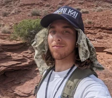
Paul Byrne
Ph.D. Student - Dinosaur Institute
University of Southern California
10:45AM - 11:00AM
Coffee break
15 minutes
11:00AM - 11:45AM
Deep Learning Automated Image Segmentation - No expertise required (Room ZHS 265)
45 minutes
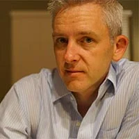
Dr. Mike Marsh
Director of Product Management, Dragonfly
Object Research Systems, Inc.
11:45AM - 12:30AM
Ion exchange processes and crystal imaging of Pb sequestration in clinoptilolite (Room ZHS 265)
45 minutes
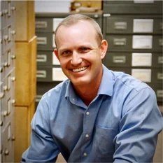
Dr. Aaron Celestian
Curator, Natural History Museum & Adjunct Professor
University of Southern California
12:30AM - 1:30AM
Lunch break
60 minutes
Workshop and tour (total 2 hours)
1:30AM - 3:30AM
CT data analysis
30 min
Aya Takase
Head of Global Marketing Communications
Rigaku
CT data collection
30 min
Angela Criswell
Director of X-ray Imaging
Rigaku
Tour at the Natural History Museum of Los Angeles County
30 min
Dr. Aaron Celestian
Curator, Natural History Museum & Adjunct Professor
University of Southern California

Contact Us
Whether you're interested in getting a quote, want a demo, need technical support, or simply have a question, we're here to help.
Subscribe to the X-ray CT Email Updates newsletter
Stay up to date with CT news and upcoming events and never miss an opportunity to learn new analysis techniques and improve your skills.