CORAL SURFACE AREA AND POROSITY ANALYSIS

About the sample: Corals

Corals are marine invertebrates. They typically form colonies of many individual polyps. Coral species include the important reef builders that inhabit tropical oceans and secrete calcium carbonate to form a hard skeleton. Scleractinia, also called stony corals, are practically the "engineers" of the coral reef ecosystems.

The shape-related parameters of coral skeletons, such as surface area, porosity, growth rate, and branching structure, represent major reference points for their physiology and the environment they were found in, and their role as ecosystem engineers (Wild et al., (2004), Nature, 428:66-70). However, even a parameter as simple as the surface area is not necessarily easy to measure because of the corals' characteristic complex shape. The traditional methods involve wrapping a sample with a foil, dipping the sample in wax, or approximating the shape of the sample with cylinders to measure the surface area. The first two are damaging to corals and cannot be used on live samples, and none of these methods is very accurate.

More recently, non-destructive X-ray CT (computed tomography) techniques have been used to characterize corals. X-ray CT can provide accurate surface area, porosity, and branching structure. (See Laforsch et al., (2008), Coral Reefs and Kruszyński et al., (2007), Coral Reefs)

Analysis procedure

  1. In this example, an about one-inch size coral skeleton was scanned using a micro-CT scanner, CT Lab HX.
  2. The CT image was segmented into the calcium carbonate skeleton and pores using thresholding segmentation.
  3. The surface area, porosity, pore size, and pore shape, were analyzed using the segmented result. Branch angles were also measured.

1. CT scan

An about one-inch size coral skeleton was scanned to produce a 3D grayscale CT image.

coral skeleton

The 3D rendered volume and a cross-section of the CT image are shown. In the CT cross-section, the calcium carbonate appears bright while the air appears dark. You can see the detailed structure of the skeleton consisting of arrays of submillimeter size pores.

CT scan of coral skeleton
CT cross-section of coral skeleton

2. Image segmentation

Segmentation for this sample was straightforward because the skeleton and the air have very high contrast. The two phases were segmented by thresholding the gray level.

coral skeleton calcium carbonate and air segmentation

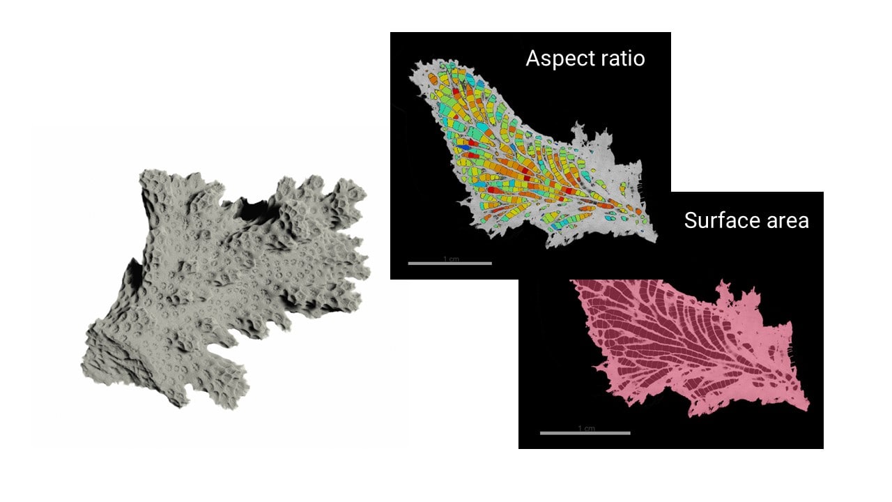
3. Surface area, pore, and branch angle analysis

Once the CT image is segmented, various shape and morphology parameters can be calculated to characterize coral samples.

From the skeleton and pore segmentation results, the porosity was calculated as 22.4%.

To calculate the outer surface area excluding the internal surfaces, we closed all pores and generated a solid coral region of interest as shown in the next figure. The surface area was calculated as 4.45 cubic centimeters.

coral surface area calculation

The next figure shows the results of the pore size analysis. The individual pores were separated by using the watershed transformation and the volume of each pore was calculated.

The pore size was relatively uniform, ranging from 0.1 to 7E8 cubic microns with a mean value of 9E7 cubic microns. The thick center part of the sample exhibited larger pores while they became smaller towards the surface or the end of the "branches."

coral pore size distribution

The next figure shows the results of the aspect ratio analysis of the pores. There is no obvious relationship between the aspect ratio and the location of the pores. The aspect ratio distribution showed a peak around 0.6, and the mean value was 0.49.

coral pore aspect ratio calculation

Lastly, a few major branches were selected and their relative angles were measured. The 3D image of the coral skeleton allows us to look inside of the skeleton on an arbitrary plane to measure the angles between branches.

coral branch angles