AMMONITE FOSSIL SCAN
About the sample: Ammonite Fossil
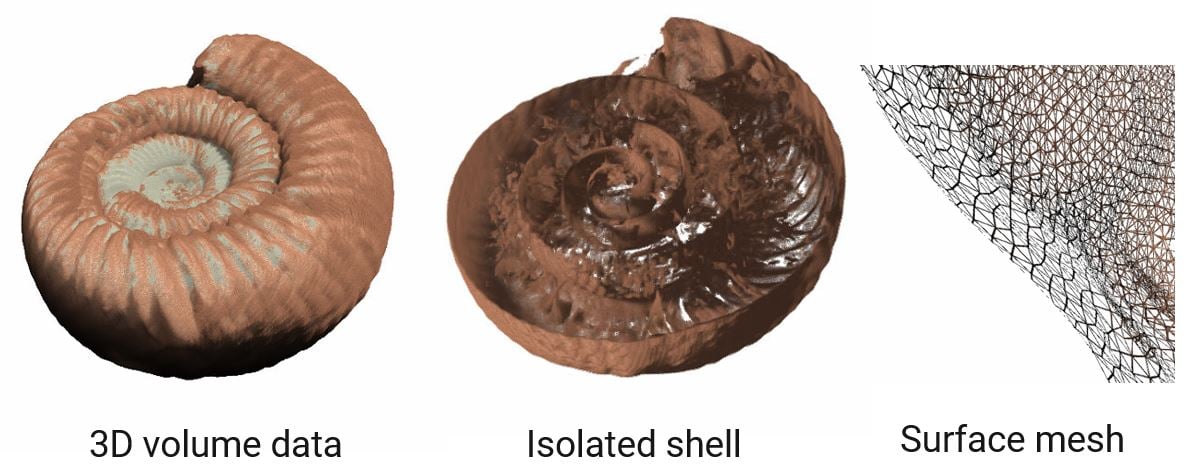
Ammonites (ammonoids) are a group of extinct cephalopods and are often found as fossils in marine rocks. Ammonites can link rock layers to specific geologic time periods based on their particular species or genus. An ammonite fossil is not the original ammonites themselves but more like its replica made of rocks. By using X-ray CT (computed tomography), we can visualize the ammonite shell including its internal structure, create a surface mesh to conduct further analyses, or make a 3D printed model.
Analysis procedure
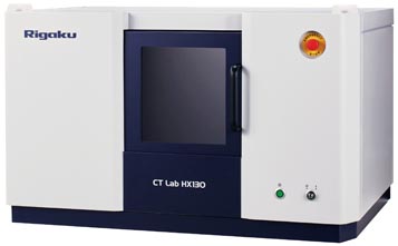
- In this example, an ammonite fossil (Perisphinctes) was scanned using a micro-CT scanner, CT Lab HX.
- The ammonite shell was segmented using the deep learning segmentation technique.
- The segmented shell was converted into a surface mesh.
1. CT scan
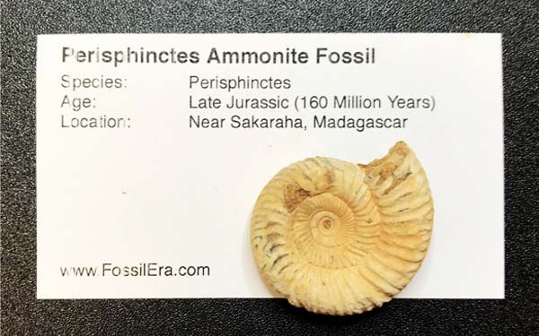
A roughly one inch size ammonite fossil (Perisphinctes) was scanned to produce the 3D grayscale CT image.
2. Image segmentation
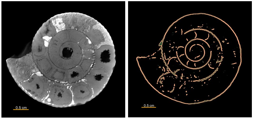
The gray level in CT data (left) represents the relative density. The ammonite shell appears slightly darker than that of the minerals filling the surrounding space.
Because the shell gray level and the air gray level (black) are very close, deep learning segmentation was employed to separate the shell structure (right).
3. Shell surface mesh
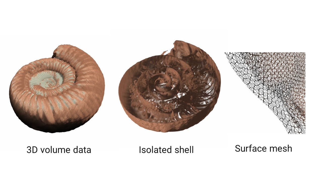
The segmented shell ROI (region of interest) was converted into a surface mesh. Once a CT image is segmented into ROIs, a ROI can be converted into a surface mesh or a volume mesh. A surface mesh can be used for dimensional analysis and 3D printing. A volume mesh can be used for various finite element analyses.
Note: You can purchase those fossils from FossilEra.
More Geological Application Examples
Watch an on-demand webinar about X-ray CT geological applications.
Sandstone grain size analysis
Application Note
Sandstone pore network analysis
Application Note & Video
Ammonite fossil scan
Application Note
Carbonate porosity analysis segmentation comparison
Application Note
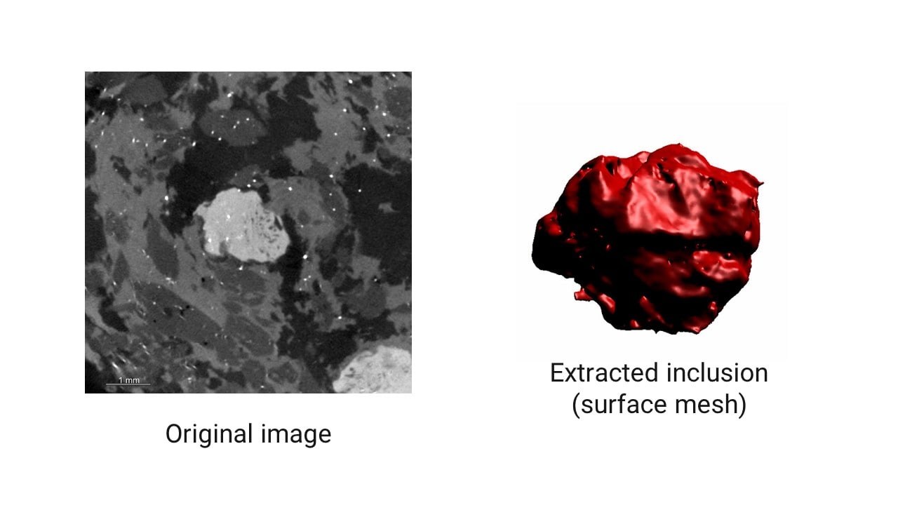
Granite phase fraction analysis
Application Note
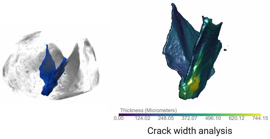
Rock crack analysis
Application Note
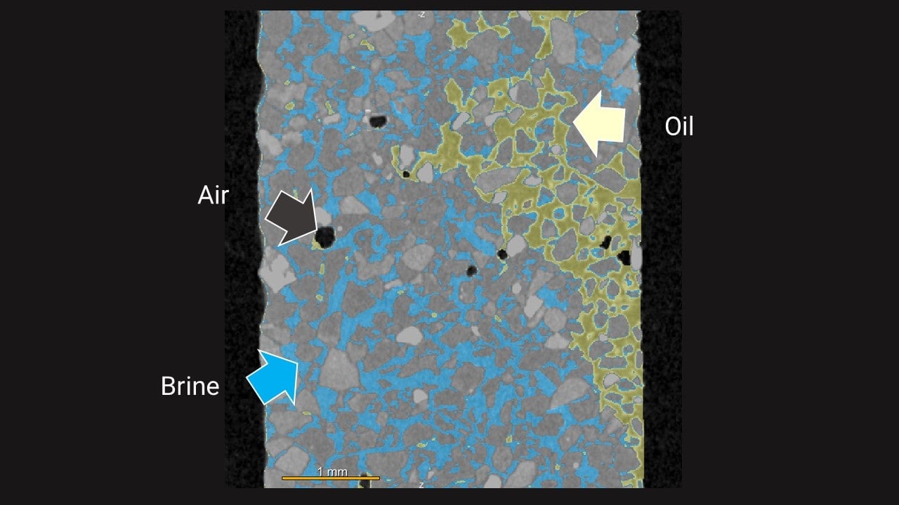
Sand oil and brine segmentation
Application Note
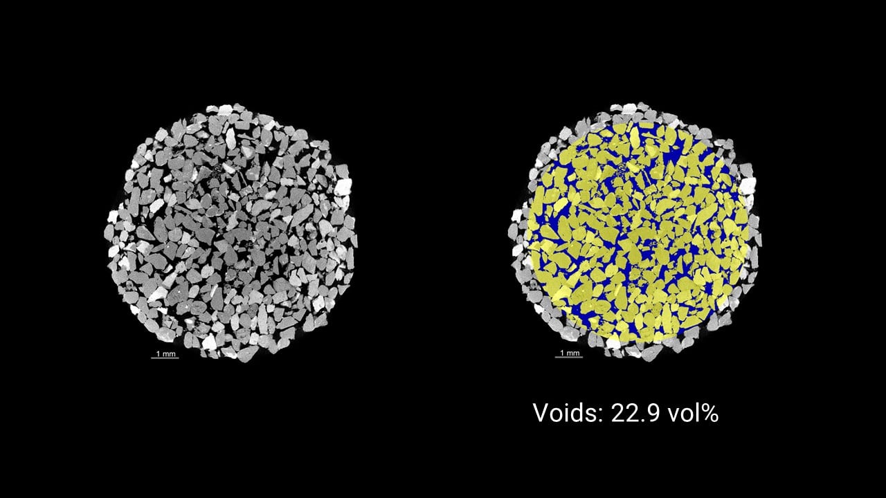
Sandstone porosity comparison
Application Note


