


Buying a scientific instrument can be a confusing and intimidating process because they are expensive, and many technical aspects are involved in selecting the right tool. While working with customers in the X-ray CT (computed tomography) industry for almost twenty years, I learned what confuses them and makes this buying process difficult. Buying a new CT scanner should be exciting, not confusing, intimidating, or overwhelming. In this blog article, I am going to answer the following questions and hope to help you enjoy your X-ray CT shopping experience:
- What is an X-ray CT scanner?
- When is X-ray CT useful?
- What types of X-ray CT scanners are available?
- How do I choose the right CT scanner?
1. What is an X-ray CT scanner
X-ray CT is an imaging technique that can non-destructively scan the three-dimensional density distribution of an object and reveal internal structures. You might be more familiar with the medical CT scanners that can reveal bones and organs in our bodies. The principles of medical and industrial CT scanners are essentially the same. However, while the resolution of the medical CT scanners is in the submillimeter to millimeter order, micro-CT and nano-CT scanners can achieve micrometer to tens to hundreds nanometer order resolution. You can use this technique to study anything from composite fiber orientations, plant phenotypes, bone structures, sandstone grain sizes, or void and crack distribution in plastic and metal parts.
Read: What is micro-CT?
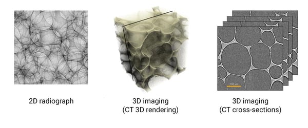
If you are already using radiography (two-dimensional X-ray imaging), you might be wondering, "what's the difference between 2D radiography and CT?" The example below shows a 2D radiograph and CT image (3D rendering and cross-sections) of a foam. All of the pores in the foam are projected on top of each other in the 2D radiograph. It makes it impossible to analyze individual pores. Meanwhile, the CT images show the foam as it is. You can see individual pores and analyze their shapes and sizes with no problem. In other words, CT removes all the difficulty of interpreting 2D radiographs.
2. When is X-ray CT useful?
If you need to image an internal structure of something non-destructively, X-ray CT can be useful. For example, you can see cracks and aggregates in a drug tablet. Or you can inspect 3D printed objects with intricate internal structures. If you use electron or optical microscopes and need an alternative or complementary technique that provides 3D images instead of 2D, or you want to avoid sample sectioning, drying, staying, coating, etc., X-ray CT can be useful. (See application examples in various industries.)
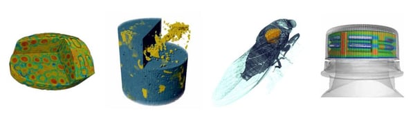 Application examples: from the left – drug table, calcite drill core, insect, bottle cap
But X-ray CT has its limitations, and it is important to know its pros and cons:
Pros
- Non-destructive
- Three-dimensional information
- Virtually no special sample preparation
Cons
- Limited sample size and density
- Limited resolution
- No elemental information
X-ray CT is an X-ray absorption contrast imaging. So X-rays need to get through the sample to generate a useful image. If the sample is high-density (heavy-absorbing) or very thick, X-rays will struggle to get through. An entire car engine is about the limit of the general industrial X-ray CT scan technique to give you an idea.
The resolution of most laboratory X-ray CT scanners is limited to microns to submicron. By using special optics, such as Fresnel zone plates, often combined with synchrotron radiation, you can achieve about 20 nm resolution. You need to switch to electron microscopes to achieve nm resolution.
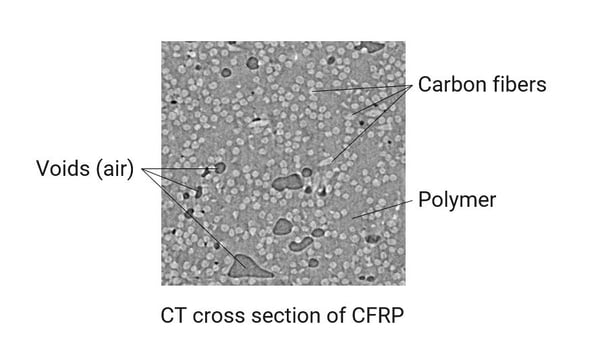
Also, it is worth noting that X-ray CT only gives us relative density information. So you cannot identify the chemical compositions or elemental makeup of the sample. If you know what types of materials are included in the sample, then you can match each gray level in the CT image to a corresponding material. For example, in CFRP (carbon fiber reinforced polymer), the lightest gray is carbon fibers, the middle gray is the binding polymer, and the dark gray is air.
3. What types of X-ray CT scanners are available?
An X-ray CT scanner consists of an X-ray source, detector, sample stage, and radiation enclosure. Depending on the required voltage and X-ray focus size, different X-ray sources are used, including reflection or transmission microfocus sources, rotating anode microfocus sources, or synchrotron radiation. Generally speaking, high voltage means high energy for X-ray sources. For micron to mm resolution systems, a flat panel or linear detector is used. Standard micro-CT and high voltage industrial CT scanners use either the cone beam or fan beam geometry, and the X-ray divergence magnifies the sample images. For higher resolution, the parallel beam geometry with an optical lens or Fresnel zone plates are used.
High-voltage industrial CT scanners tend to be on the expensive side, about half a million to multi-million dollars, depending on the required voltage and level of automation. This is because of the high price of a high-voltage X-ray source and a large sample stage and radiation enclosure to accommodate large and heavy samples. Standard micro-CT scanners are one of the most affordable. For a research-grade micro-CT scanner, the price ranges anywhere from mid-200K to half a million dollars. Compact benchtop-type scanners are also available in this class. Nano-CT scanners are relatively expensive and range from about half a million to two million dollars, depending on the required resolution. There are many CT manufacturers. For example, Rigaku makes nano- and standard micro-CT scanners, and companies like Yxylon and Nikon make high voltage industrial CT scanners.

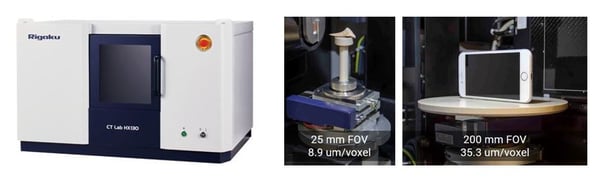 A benchtop micro-CT scanner: CT Lab HX exterior (left) and sample stage (right)
Read: How Much Does a Micro CT Scanner Cost?
4. How do I choose the right CT scanner?
The right CT scanner type depends on the sample type and size of the features you want to image. There are three major specifications to consider: X-ray energy (or voltage), field of view (FOV), and resolution. The FOV is the area you can scan, and it is normally similar to the sample size. The table below summarizes the sample factors and X-ray CT specifications:
|
Sample factors |
CT specifications |
What does this mean? |
|
Density |
X-ray energy |
For example, a piece of 30 mm thick steel needs a high-energy/high-voltage X-ray source, 200 kV or above. Small to medium-sized aluminum, ceramics, plastic, etc., need only a standard voltage X-ray source, 60-160 kV. Organic materials can benefit from low-energy (5-20 keV) nano-CT. |
|
Sample size |
FOV X-ray energy |
FOV should cover the area you want to scan. It should be similar to the sample size. Large samples need a high-energy/high-voltage X-ray source, 200 kV or above. Smaller samples need only a standard voltage X-ray source, 60-160 kV. |
|
Resolution |
Spatial resolution |
The spatial resolution needs to be equal to or smaller than the size of the features you want to see. |
The optimum X-ray energy depends on the sample density and thickness X-rays need to get through. Generally speaking, for larger or heavier samples, such as steel or copper thicker than a few mm, you will need high-energy (100 - 400 keV). Small to medium-sized (5~200 mm) plastics, polymers, composites, wood, etc., or small (a few mm) and light metals (aluminum for example), medium-energy X-rays (20 - 100 keV) work well. For relatively small (1~10 mm) organic materials, foams, etc., low-energy X-rays (5 - 20 keV) work best.
For the FOV and resolution, keep in mind that the spatial resolution is limited to about 1/1000 of the FOV. This is because the number of pixels on commonly used X-ray detectors is limited. (Learn more about resolution. à this will link to a resolution blog article.)
Regardless of the technical requirements, the instrument size and price are always important. If your lab space is limited, a benchtop system can save a lot of space. Always check the dimensions and weight of the system. CT scanners can be heavier than they look because of the thick metal radiation shield stopping the high-energy X-rays. For the price, as summarized in the previous section, the standard micro-CT scanners are the most affordable. The higher X-ray energy or resolution is required, the higher the price becomes.
Lastly, you should always ask the manufacturer to scan a few representative samples of yours to make sure that you are happy with the image quality. If you are new to CT and considering purchasing your first CT scanner, I would also recommend that you visit the manufacturers either in person or via a video conference to have a live demonstration to really understand what is involved in running the instrument.
If you have questions or would like to try one of Rigaku's CT scanners, talk to one of our CT experts by clicking the “TALK TO AN EXPERT” button at the right top of the page or contact us at imaging@rigaku.com.