


Industrial CT and medical CT both use the X-ray CT (computed tomography) technique to visualize the internal structures of an object non-destructively. They share the same basic principles.
You might ask, “What are the differences between these two types of CT scanners?” “Can we use a medical CT scanner for non-medical applications?” “Which one is more technologically advanced?” These are all good questions, and I will answer them in this article.
My experience is mainly with industrial—especially micro and submicron—CT scanners. While these CT scanners are built for industrial inspections and materials research and have unique advantages over medical CT scanners, I have always been fascinated with the advances made in the medical CT industry. These advances are usually modified and applied to industrial CT scanners later.
In this article, we will look at the differences between the two types of CT scanners from several perspectives:
- What are the differences between industrial and medical CT scanners?
- Application differences - Do we ever use a medical CT scanner for non-medical purposes?
- Software differences - Do we process the images differently?
- Industry/market differences - Which one is leading innovation?
What are the differences between industrial and medical CT scanners?
There are several critical differences between industrial and medical CT scanners. These differences come from what is required for industrial inspection and materials research (industrial CT) versus medical diagnostics (medical CT).
The table below summarizes the differences:
|
Industrial CT |
Medical CT |
|
|
Requirements |
||
|
Allowed dosage |
Minimum to no limit |
Limited |
|
Sample/patient position |
Horizontal or vertical with rotation |
Horizontal with translation |
|
Sample size |
1 - 300 mm |
1 - 2 m |
|
Resolution |
0.5 - 100 microns |
~ 0.5 mm |
|
Grayscale |
Relative scale |
Normalized scale |
|
Scan object |
Varies widely (organics, plastics, ceramics, metals, etc.) |
Human body |
|
How to meet these requirements |
||
|
Scan geometry |
Sample rotation with a fixed X-ray source and detector |
X-ray source and detector rotation (gantry system) with the patient lying stationary on a bed |
|
X-ray geometry |
Cone or parallel beam geometry, or their combination |
Fan or cone beam geometry |
|
Scan time |
A few seconds to hours; typically 30 - 120 min |
~ 5 seconds |
|
X-ray voltage |
90 kV - 9 MV; typically 100 - 225 kV |
120 - 140 kV |
|
Number of slices |
256 - 3000 |
6 - 128 |
Industrial CT
Resolution
Industrial CT scanners—often referred to as mico-CT scanners—are used for samples ranging from 1 mm to usually about 300 mm. CT scans help detect small defects in machined, printed, or assembled products for inspection and failure analyses. CT scans are also used to see detailed micron-to-submicron structures of foams, composites, etc., for materials research purposes. In both cases, relatively high spatial resolution is required, ranging from submicron to microns, while submillimeter resolution suffices for medical CT.
To achieve high resolution, you need a smaller X-ray focus, a small detector pixel, and a high magnification factor. All of these factors affect the design of a CT scanner.
Achieving high resolution requires a longer scan time because the higher the magnification factor and the smaller the voxel size, the fewer X-ray photons go into one pixel of a detector. With the limited X-ray power and detector size available today, this translates to lower X-ray intensity. You need to increase the scan time to compensate for the intensity loss and achieve good image quality with a high signal-to-noise ratio.
This can be a problem for medical CT because you want to avoid exposing a patient to radiation for a long time, but it is not a problem for industrial CT.
Geometry
When achieving submicron to a few microns resolution, even a small misalignment of the X-ray source, rotation center, or detector center can blur the resulting CT images. So, it makes sense to rotate a small sample on a precision rotation stage instead of using a gantry system to spin a heavy X-ray source and a detector around a sample. This is why micro-CT scanners commonly use horizontal geometry with sample rotation.
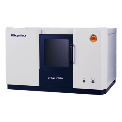
Example industrial CT scanner: Benchtop industrial micro-CT scanner
Meanwhile, medical CT scanners use the gantry system and spin an X-ray source and detectors around a patient at high speed because rotating a patient at high speed is not practical.
Horizontal gantry systems are used occasionally for in-situ or operando industrial CT scans to keep the sample stationary and make it easy to change the sample environment during measurements.
X-ray voltage, contrast, and grayscale
For industrial and materials research applications, the scan objects can be anything from organic materials—such as tissue samples, plants, polymers, and plastics—to metals. This requires the X-ray voltage to be adjustable to achieve good contrast. Also, since we usually know what types of materials are in the sample, we can guess what is what from the relative gray levels. The gray levels need to be somewhat absolute for medical CT scanners, as we discuss next.
Medical CT
Radiation dosage
Limiting the radiation dosage is one of the most important things to consider when designing a medical CT scanner. This limits the scan time. The resolution doesn’t need to be high; submillimeter usually suffices.
Geometry
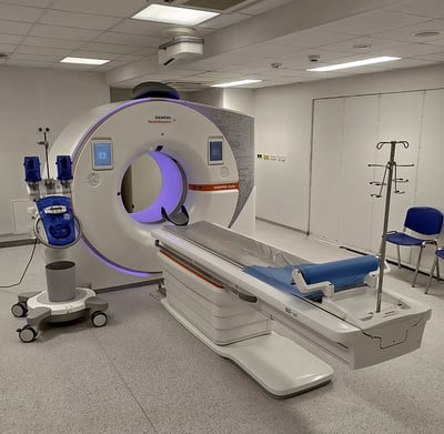
Modern CT scanner located at the Lochotín University Hospital in Pilsen, Czech Republic.
Letting the patient lie on the table during a scan is also important. For this purpose, medical CT scanners use the gantry system and spin an X-ray source and detectors around a patient at high speed while the patient moves through the X-ray beam path. You can see how fast the gantry spins during a CT scan in this video:
Radiographer Films Inside of a CT scanner spinning at full speed.
You can see why spinning the patient is not a good idea for medical CT scanners.
X-ray voltage, contrast, and grayscale
Because the scanned object is always a human body, we can optimize the X-ray voltage for it. At the optimized voltage for a specific scanner, the gray levels for air and water are measured and used to normalize the measured gray levels on patients.
The normalized values are expressed in the Hounsfield unit, also referred to as the CT unit ranging from -1000 (air) to about 2000 (high-density bones such as cochlea). Average bones are around 1000. This normalization helps radiologists identify tissues, bones, and their abnormalities in CT images.
Application differences - Do we ever use a medical CT scanner for non-medical purposes?
As we reviewed in the previous section, the differences between industrial and medical CT scanners come from the differences in scanned objects and required sample settings and image qualities.
Do we ever use a medical CT scanner for non-medical purposes?
Yes, there are several materials research areas in which it makes sense to use medical CT scanners. These include imaging of drill cores for digital rock physics and anything too long to scan on conventional industrial CT scanners, like tree trunks.
If you are wondering how the data compare, you can see a comparison of industrial and medical CT scanners in this case study by du Plessis et al. The authors compared data on a steel turbine blade, a titanium casting, a concrete cylinder with aggregates and pores, and a concrete block with metal fiber reinforcement.
A medical CT scanner might be the only one you can access at your facility. If that’s the case, there is nothing wrong with scanning your sample using a medical CT scanner. You don’t need to purchase a separate industry CT scanner if you can get enough resolution and good image quality for the required analyses.
Software differences - Do we process the images differently?
We have focused on the hardware differences so far, but is the software for image processing different?
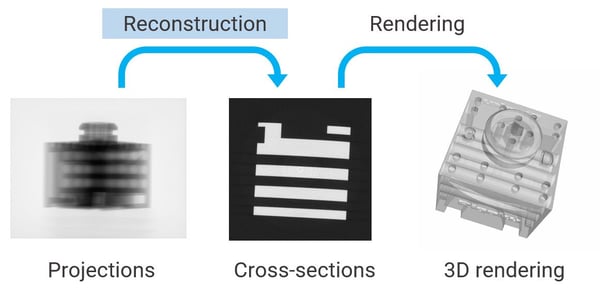
Both types of CT scanners usually use the filtered back projection method to reconstruct the CT images from the 2D radiographs or projections. However, there are some differences:
- While most industrial CT scanners use cone beam geometry and axial scans, many modern medical CT scanners use fan beam geometry and helical (spiral) scans. The exact calculation involved in reconstruction is different between the two. (To learn more about different scan modes, refer to “Illustrated comparison of CT Scan Modes [axial, helical, wide-cone axial, low pitch, high pitch] for Radiologic Technologists” by Dr. Brian Nett.)
- A relative grayscale is commonly used in industrial CT, while the normalized scale, the Hounsfield unit, is used in medical CT. Medical CT requires an additional step to normalize the grayscale after reconstruction.
Reconstruction is a process to convert a set of 2D radiographs (projections) to a 3D volume consisting of CT slices. This should not be confused with a 3D rendering of volumetric data.
To learn more, watch the workshop "Demystifying Reconstruction Using ImageJ"
After reconstructing CT images, image segmentation and quantitative analyses follow for industrial CT. The analyses include simple phase volume fraction, particle size distribution, and dimensional analyses. For medical CT, radiologists read the CT images and diagnose the injuries and diseases. Quantitative analyses are sometimes used for medical purposes, such as a cardiac CT calcium score, also known as a coronary calcium scan.
As discussed in the next section, reducing radiation dosage remains an objective of medical CT scanner development because a lower dosage is better for the patients. This need has driven the development of new reconstruction algorithms that can help reduce the dosage. Industrial CT scanners benefit from the same algorithms when increased scan speed is needed for in-situ and operando measurements.
Industry/market differences - Which one is leading innovation?
The first commercial CT scanner was developed for medical use in the early 1970s. Although industrial and scientific research use of CT has become more common in the last few decades, the medical CT industry has led many technological advancements over the years. It makes sense, considering the market size difference. The medical and industrial CT market sizes in 2020 are estimated to be $5.6B and $440M, respectively.
The primary requirement in medical CT is improving the image quality and accuracy of diagnoses while reducing the radiation dosage and scan time. Iterative reconstruction algorithms and the use of deep learning in the reconstruction process advanced enough to see some real-world applications in the last ten years.
For example, over 17,000 papers have been published about deep learning techniques developed or tested for dose reduction in the last ten years. Both techniques help reduce the amount of required projection data; that is, the radiation dosage, to reconstruct tomographic images without sacrificing image quality.
Dual-energy and spectrum CT are also relatively new. They help distinguish tissues with similar densities. They also help doctors avoid scanning patients twice to gain a different view or additional information.
Photon counting CT (PCCT) has attracted attention recently, and many believe it will be a hot topic in the coming years. Photon counting detectors are not new to X-ray analytical instruments such as diffractometers, but they are yet to be utilized for medical CT scanners. PCCT has the potential to visualize smaller details of organ structures with more precise tissue characterization using spectral CT capabilities compared to traditional CT while reducing the dosage.
Lastly, artificial intelligence (AI) is increasingly applied not only to the reconstruction of CT images but also to their interpretation to help radiologists diagnose injuries and diseases faster and more accurately. The study and use of AI accelerated during the COVID-19 pandemic, and many promising results were reported.
The industrial CT market might be ten times smaller than medical CT, thus having ten times fewer development resources, but many of the technological advancements made in the medical field can be and have been applied to industrial CT. I look forward to seeing more new technologies transferred from medical to industrial CT.
Which one should I use?
As we reviewed above, industrial and medical CT scanners have a lot in common, yet there are differences that can be critical to the success of your research.
If you have only access to medical CT scanners and have been wondering if that will work for your industrial or materials research project, I hope this article helped you answer that question.
If you are not sure which type of CT scanner is suitable for your analysis needs, our team of CT experts can help you figure out the best fit. You can talk to one of our CT experts by clicking the “Talk to an expert” button at the top right of the page or sending us a message at imaging@rigaku.com.